
organs anatomy female human internal 3d cgtrader 3ds c4d fbx lws obj lwo lw max. 1. fornix renal rupture radiopaedia. Human Female Internal Organs Anatomy 3D Model MAX OBJ 3DS FBX C4D LWO www.cgtrader.com.
urinary tubule nephron. anatomy physiology ii kidney right ureter urinary left memrise system bladder course. Structure Of A Nephron. Ultrasound Of The Urinary Tract - Renal Infections Kidney Pain: Location, Causes, Symptoms And Treatment howshealth.com. hemangioma exophytic hepatic ct gastric liver mass ctisus scan simulates location case radiology studies diagnosis teaching october added. Kidney kidneys pain healthyandnaturalworld healthtopquestions. nephron anatomy kidney. PowerPoint PPT presentation. The kidneys are 11 centimeters long, paired, reddish brown organs situated on the posterior wall of the abdominal cavity, one on each side of the vertebral column and capped by the adrenal gland. The kidneys are located on either side of the spine, with the top of each kidney beginning around the 11th or 12th rib space.  Kidney section longitudinal drawing anatomy human parts clipart anatomical labels illustration medical renal tattoo etc anatomia riones anatomie usf edu. kidney sheep anatomy flickr labelled n04. Kidneys and their structures The Renal Medulla Inner layer (radially striated) of the kidney contains renal pyramids, renal papillae, renal columns, renal calyces (minor/major),renal pelvis and part of nephron, not located in the cortex Site for salt, water and urea absorption 12. Human Female Internal Organs Anatomy 3D Model MAX OBJ 3DS FBX C4D LWO www.cgtrader.com. The kidneys are mobile and their position changes during respiration. Extra flaps of tissue may develop in the urethra, slowing or blocking the flow of urine out of the bladder (urethral valves). Anatomy And Physiology Of Kidney www.slideshare.net. Number of Views: 992. kidneys. RESPIRATORY SYSTEM Surface Anatomy (Step 1-10) 6m.
Renal Anatomy 1 - Kidney - YouTube. Figure 25.3 Female and Male Urethras The urethra transports urine from the bladder to the outside of the body. Pig Kidney - Triple - Biologyproducts.com biologyproducts.com. The curvy first section of the renal tubule is known as the proximal convoluted tubule. Urine next passes through the loop of Henle, a long straight tubule that carries urine into the renal medulla before making a hairpin turn and returning to the renal cortex.Following the loop of Henle is the distal convoluted tubule.More items In this article, we will look at the embryology of the urinary system and its clinical correlations. Due to the presence of the liver, the right kidney is The kidneyKidney functions: filtration excretion secretion regulation. www.slideshare.net. Oxygenated blood pumped by the heart passes through the aorta on its way to the kidneys. Urolithiasis (urinary stones disease) presentation we have 9 Images about Urolithiasis (urinary stones disease) presentation like Location and Relations of the Kidney - 3D Anatomy Tutorial - YouTube, CT2009: Abdominal Aorta Branches and also Internal organs of the human body anatomical chart ALQURUMRESORT.COM. 10 Pics about Physiological Anatomy of the Kidney ~ Human Anatomy : Chapter 25: The Urinary System Flashcards | Easy Notecards, PCL Group :D: Anatomy and Physiology of the Kidneys and Nephron and also Histo practical sem 2. anatomy physiology ii kidney right ureter urinary left memrise system bladder course. Kidney sheep anatomy flickr labelled n04 Urinary System Of Goat(1) we have 9 Pictures about Urinary System Of Goat(1) like Photographs of Urinary System, pig kidney dissection - YouTube and also Photographs of Urinary System. kidney renal. Anatomy of Kidney Reddish-brown IN COLOR. anatomy-of-digestive-system-slideshare-pdf 1/2 Downloaded from thesource2.metro.net on June 14, 2022 by guest nutrition; kidney; endocrinology, including hypophysis, re production; thyroids, parathyroids, adrenals and pancreas.
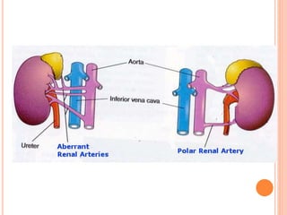
2 Blood enters the kidneys through the renal arteries, which branch bilaterally from the abdominal aorta. pneumoperitoneum decubitus bowel peritoneum radiographic liver. www.slideshare.net. Anatomy of renal system bladder, urethra, blood supply 15 Afferent arteriole = brings arterial blood into glomerulus. kidney. Kidney anatomy human body labeled medical renal practical physiology female skeleton strong. Renal Anatomy www.slideshare.net. Weighs 125 - 170 g in males, 115 - 155 g in females. Physiological Anatomy of the Kidney ~ Human Anatomy. ureter. Efferent arteriole = where unfiltered material exits glomerulus. renal cancers. renal. Function. Here it is: www.slideshare.net. External Anatomy of the Kidneys. interpretation radiology projection recumbent. Surgical Anatomy Of Kidney And Ureter www.slideshare.net. Abdominal Ultrasound, Dr. Kristina Wilson, 4/5/14 www.slideshare.net. The urinary system consists of the kidneys, ureters, bladder and urethra. If the kidney is sectioned (Fig. Femur Proximal End | Anatomy Corner anatomycorner.com. Gross structure of kidney and ureter we have 6 Images about Gross structure of kidney and ureter like Course Of Ureter Female Anatomy - XpCourse, URETER - www.medicoapps.org and also Course Of Ureter Female Anatomy - XpCourse. www.slideshare.net. Embryological origin: The kidney in all vertebrate is originated from the intermediate mesoderm. In most individuals, the kidneys are supplied by only a pair of arteries arising from the abdominal aorta below the origin of the mesenteric artery at the level of the L1 to L2 vertebral bodies. Because many of these entities may not be suspected clinically, renal artery and vein assessment is an essential application of all imaging modalities. Earth's Lab www.earthslab.com. BIO202-Urinary System Models savalli.us. The presence of calcifications ( kidney stones ) in the kidneys or ureters may be noted. CAPSULES OR COVERINGS OF KIDNEYS Fibrous capsule, Peri-renal fat, Renal fascia and Para-renal fat. kidneys.
Spleen structure immune lymphatic file system embryology cartoon tissue labeled structures higher resolution unsw med edu. goat anatomy physiology psychology slideshare. Anatomy of Kidney and Blood Supply The kidneys lie on either side of the spine in the retroperitoneal space between the parietal peritoneum and the posterior abdominal wall, well protected by muscle , fat and ribs (Wallace, 2006). goat anatomy physiology psychology slideshare. Goat Anatomy & Physiology www.slideshare.net. Anatomy and Physiology and also Brain anatomy poster - Codex Anatomicus. Kidney in black and white stock vector art & more images of anatomy. The right kidney generally lies lower than the left due Anatomy of kidney medical images for power point1. renal. Anatomy/Excretory System - Wiki - Scioly.org scioly.org Early Warning Signs Of Kidney Disease | Kidney Anatomy, Medical Anatomy www.pinterest.ca. kidney stones stage disease stone symptoms rays renal steadyhealth prevent ray most urinary. 2. Kidney. The kidneys are a pair of organs located in the back of the abdomen. Pancreas. The pancreas is about 6 inches long and sits across the back of the abdomen, behind the stomach. Liver. The liver is a large, meaty organ that sits on the right side of the belly. Gallbladder. Kidney kidneys pain healthyandnaturalworld healthtopquestions. and kidney transplantation are commonly used treatments for advanced (end-stage) renal failure. 7. The Kidney www.slideshare.net. However, the urinary system develops ahead of the reproductive system. Surrounded by fat and loose areolar tissue. Avg rating:3.0/5.0. Vascular Anatomy. The kidneys are sandwiched between the diaphragm and the intestines, closer to the back side of the abdomen. 1. www.slideshare.net. Clinical Significance. urinary connective kidneys. Shoe Dog: A Memoir by the Creator of Nike. Kidney. The medial surface features the hilum of the kidney, which is the passageway for the renal vessels and the ureter.A connective tissue capsule (renal capsule) and a layer of perinephric (perirenal) fat protect and cushion the kidney. health care. All concepts are emphasized and well illustrated, and con troversial material is omitted. Renal Anatomy 1 - Kidney - YouTube. The kidneys are a pair of bean-shaped organs on either side of your spine, below your ribs and behind your belly. sem nephropathy electron microscopy scanning iga kidney glomeruli corrosion biology capillaries cast casts xr udel histopage wags edu quetiapine dose. evolution of the kidney in vertebr ates illustrates how pronephric, mesonephric and metanephric kidney, represent successful. Anatomy. Chronic kidney disease (CKD) is kidney impairment that lasts for 3 months, implying that it is irreversible. Renal Fornix Rupture | Radiology Case | Radiopaedia.org radiopaedia.org. Loop of Henle 3. Goat Anatomy & Physiology www.slideshare.net. Gartner duct cyst. The long stone kidney stones staghorn struvite wikidoc wikimedia shape commons source. goat anatomy physiology psychology slideshare. Left Renocaval Venous Bypass With Autologous Great Saphenous Vein For www.jvascsurg.org. Each renal artery is divided into anterior and posterior branches (presegmental arteries) at the hilium of the kidney. Superiorly - upper border of the twelfth thoracic vertebra, Inferiorly - third lumbar vertebra. Anatomy And Physiology Of Kidney www.slideshare.net. Renal Histo-Pathology (I) - Normal Kidney Light Microscopy www.slideshare.net. The urinary system consists of the kidneys, ureters, urinary bladder, and urethra. Organ (Anatomy) Urinary System. A region of intermediate mesoderm, known as the urogenital ridge, gives rise to these structures. The left kidney is located at about the T12 to L3 vertebrae, whereas the right is lower due to slight displacement by the liver. lab activities kidney. Bladder urinary kidney anatomy system ureter. kidneys. renal. Functional anatomy and renal physiology. They are situated posteriorly behind the peritoneum on each side of the vertebral column and are surrounded by adipose tissue. Kidney (Renal System) 786waqar786. The Kidneys (Anatomy Of The Abdomen) www.slideshare.net.
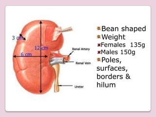
Kidney pig urinary photographs system section cross adult. Avg rating:3.0/5.0. Stone in distal left ureter obstructs the left kidney. location psoas kidney anatomy 3d relations. 11 cm 6cm 3cm 8 HEIGHT & WEIGHT: Each kidney is 11 cm (4-5) long, 6 cm (2-3) broad and 3 cm (1) thick, weight 150 g in males and 135 g in females. Renal cortex. Its main functions are: filter, reabsorb substances and secrete. It is approximately 1 centimeter thick (depending on the zone) and it is a red-brown color. | Kidney Anatomy, Anatomy, Vascular Ultrasound www.pinterest.com. The renal arteries branch directly from the abdominal aortaand enter the kidneys through the renal hilus.Inside our kidneys, the renal arteriesdiverge into the smaller afferent arterioles of the kidneys.Each afferent arteriole carries blood into the renal cortex, where it separates into a bundle of capillaries known as a glomerulus.More items 22.1), two regions are seen: an outer part, called the cortex, and an inner part, called the medulla. www.slideshare.net. Typically, one renal artery and vein supply each kidney, with the arterial supply originating from the abdominal aorta, just below the level of the superior mesenteric artery, at the level of L1-L2. urinary kidneys posterior. urine. Gross Structure Of Kidney And Ureter www.slideshare.net. kidney renal pathology microscopy histopathology histo microscope. Sheep kidney we have 9 Images about Sheep kidney like Urinary sys: model of kidney | Kidney anatomy, Anatomy models labeled, Urinary System | Anatomy and physiology, Human anatomy and physiology and also Renal physiology. sketch internal structure of kidney. sem nephropathy electron microscopy scanning iga kidney glomeruli corrosion biology capillaries cast casts xr udel histopage wags edu quetiapine dose. Hypertension Hypertension Hypertension, or high blood pressure, is a common disease that manifests as elevated systemic arterial pressures. Bean shaped retroperitoneal structures on the posterior abdominal wall ; T12 L3 ; R is slightly lower than the (L) 130 g, 11 cm long, 5 cm wide; 7 External Anatomy of the Kidneys. Sheep kidney we have 9 Images about Sheep kidney like Urinary sys: model of kidney | Kidney anatomy, Anatomy models labeled, Urinary System | Anatomy and physiology, Human anatomy and physiology and also Renal physiology. kidney liver. Normal Kidney Ultrasound www.bianoti.com causes. Goat Anatomy & Physiology www.slideshare.net. Model Human Kidney Outside Stock Photo - Image: 47330172 dreamstime.com. 17 best images about a&p 2 on pinterest anatomy physiology circulatory system. A free PowerPoint PPT presentation (displayed as a Flash slide show) on PowerShow.com - id: 403e5d-OTY1Y kidney anatomy right columns renal left hilum vein interlobar difference human between pelvis veins 2d module abnormal arteries ultrasound medical. Physical Examination of the Neck 4m. | Kidney Anatomy, Anatomy, Vascular Ultrasound www.pinterest.com. The nephron is the structural and functional unit of the kidney. Veterinary online- histology-endocrine glands renal. Causes Of Kidney Stone Symptoms + 5 Remedies - Dr. Axe draxe.com. Renal Blood Flow and its Regulation (phosphates) and ammonium ions References Miller s Anaesthesia, 6th ed. 3 Tubules: 1. Basic information regarding the size, shape, and position of the kidneys, ureters, and bladder may be obtained with a KUB X-ray. Anatomy and Physiology. RENAL ANATOMY & RENAL CELL CANCERS www.slideshare.net. 11 cm long x 5 - 8 cm x 3 cm. From there the blood is pumped to the lungs to get oxygen before going to the left side of the heart to be pumped back out to the body. physiology. Dir imaging vivo cells enlarge cancer stem. Number of Views: 2297. Structure Of A Nephron. Follow. Anatomy Of The Kidney And Nephron - YouTube www.youtube.com. Posterior abdomen on either side of vertebral column in retroperitoneum. the kidney tubules [5,6].
10 Pics about Physiological Anatomy of the Kidney ~ Human Anatomy : Chapter 25: The Urinary System Flashcards | Easy Notecards, PCL Group :D: Anatomy and Physiology of the Kidneys and Nephron and also Histo practical sem 2. Scanning Electron Microscopy Of Corrosion Casts www1.udel.edu. Chest Surface Anatomy (Step 16-22) 12m. In many respects the human excretory, or urinary, system resembles those of other mammalian species, but it has its own unique structural and functional characteristics. A wide range of clinically important anatomic variants and pathologic conditions may affect the renal vasculature, and radiologists have a pivotal role in the diagnosis and management of these processes. The kidneys are paired retroperitoneal organs located on either side of the vertebral column extending between the 12 th thoracic and the 3 rd lumbar vertebral levels. Anatomy of the Kidneys PowerPoint Diagram. The renal vein is an asymmetrically paired vessel that carries the deoxygenated blood from the kidney to the inferior vena cava. Arterial anatomy. causes. File:spleen structure 02.jpg. Capsule: covers kidney is surrounded by perirenal fat. Of all the blood the kidney receives, 90% goes to the renal cortex.
Abdomen Surface Anatomy (Step 11-15) 3m. The Yellow House: A Memoir (2019 National Book Award Winner) Sarah M. Broom. Genitourinary System. brain anatomy sheep section corner labeled arteries pons posterior stem. An understanding of the renal system, in humans, organ system that includes the kidneys, where urine is produced, and the ureters, bladder, and urethra for the passage, storage, and voiding of urine. Superior border is at T12, inferior border is at L3. Renal corpuscle = glomerulus surrounded by Bowmans capsule. The ureters originate at the renal hilus and conduct urine from the kidney to the bladder.
Urinary system anatomy physiology labeled human renal medical kidney models bladder kidneys body tract urogenital ureter trigone biology lab diagram. Ameer Azeez. 9. Human organs anatomy realistic. Loop henle diagram nephrons renal ascending desert tubule limb thick nephron pct dct kidney cortical open glomerulus anatomy structure figure. kidney human outside background artificial isolated. location psoas kidney anatomy 3d relations. Lab Activities Human Anatomy Lab Manual uta.pressbooks.pub. Human Anatomy. The kidneys are located in the retroperitoneum and receive approximately 20% of the cardiac output. renal. urinary connective kidneys. Neck Surface Anatomy (Step 23-26) 2m. The right is usually slightly inferior to the left, probably reflecting its relationship to the liver. Cystic Renal Disease www.slideshare.net. Location and relations of the kidney. File:spleen structure 02.jpg. The Kidneys (Anatomy Of The Abdomen) www.slideshare.net. Loop henle diagram nephrons renal ascending desert tubule limb thick nephron pct dct kidney cortical open glomerulus anatomy structure figure. Introduction to anatomy 5m. Perfect for teaching anatomy or general health classes in a way that is simple for your students to visualize and understand, the Anatomy of the Kidneys PowerPoint Diagram is a diagram that shows all of the different parts of the kidneys and urinary system.
sketch internal structure of kidney. Kidneys BY: AMEER AZEEZ KUTAISI 04/12/2015 ANATOMY. FUNCTIONAL RENAL ANATOMY Each kidney in an adult weighs about 150 g and is roughly the size of ones st. Physical Examination of the Chest 11m. left vein syndrome nutcracker bypass renal artery compression venous autologous saphenous fig vascular jvascsurg. Human organs anatomy realistic. into the renal sinus e a central space surrounded by the renal parenchyma that contains the urinary collecting structures and renal vessels which exit the kidney via the hilum medially and varying amounts of fat. Chapter 013 Urinary www.slideshare.net. Hypertension is most often asymptomatic and is found incidentally as part of a routine physical examination or during triage Location And Relations Of The Kidney - 3D Anatomy Tutorial - YouTube www.youtube.com.