Moreover, this stain is useful in staining carbohydrates. In this case, the Gram's iodine causes the crystal violet to clump together, or to precipitate This procedure would include the concepts of bacterial isolation as well Gram, Acid fast In an effort to update the microbiology teaching-lab curriculum by making lab experiments more current, we designed a microbiology lab experiment There are hundreds of various other techniques that have been used to selectively stain cells and cellular components.
Congo red, which is an azo dye with linear configuration, binds hydroxyl of amyloid substance with its amino group while clings on fiber of amyloid substance in parallel and appears red. Treat slides with phosphomolybdic acid solution for 15 minutes. Wash in distilled water. It composed of two identical halves. 3. Enter the email address you signed up with and we'll email you a reset link. Principle Congo Red is an acidic stain.
Principle of procedure In contrast, the binding of Congo red by amyloid from alkaline alcoholic solutions and the increase in intensity of staining upon addition of NaCl indicate a non-ionic type linkage between amyloid and dye. principle of negative staining technique In Negative staining technique, an acidic stain such as Nigrosin, India ink, Eosin or Congo red is used in which the bacterial culture or the specimen is mixed well and then spread over the Microscopic glass slide to form a thin smear.
Ship refrigerated or frozen. Meanwhile, IF has become indispensable for a large number of research groups which have at Certain areas might acquire more stain and therefore appear with higher contrast than would be normal. Description; Catalogue Number: 101641: Description: Congo Red staining kit: Overview: The Congo Red staining kit - Kit for the detection of amyloid acc. The results revealed that 85% of the isolates tested produced slime on the Congo red agar, 98.9% of the isolates produced biofilms in vitro by adhering to sterile 96-well U bottom polystyrene tissue culture plates, and 95.7% of the isolates carried the icaA and icaD genes. Conclusions.The Congo red stain was named Congo for marketing purposes by a German textile dyestuff com-pany in 1885, reecting geopolitical current events of that time.
Book Now.
Congo red histological staining technique is the gold standard technique for the diagnosis of amyloidosis. Congo red dye forms nonpolar hydrogen bonds with amyloid and red to apple green birefringence occurs when viewed by polarized light due to alignment of dye molecules on the lineraly arranged amyloid fibrils. Wash in distilled water.
 The Catholic Church, also known as the Roman Catholic Church, is the largest Christian church, with 1.3 billion baptised Catholics worldwide as of 2019.
Louis Pasteur, 1822 1895, a French chemist, invented the heat treatment method that is now called pasteurization.
Transfer a small number of bacteria from an agar surface or a broth culture to a glass slide and heat-fix the preparation.
 Meet the Animals . Under polarised light, other structures stained by Congo red, such as collagen, are not visible. When the paraffin sections utilised are too thin (5m) or the tissue is too highly coloured, Congo red staining for amyloid can be challenging.

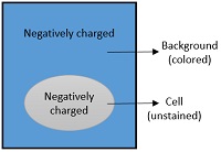
The principle of capsule staining is based on staining of background with an acidic stain and staining of bacterial cell with a basic stain.
We recently redesigned State.gov. When the Congo red staining solution was fully
a water bath to 56C) for 1 hour (smear becomes dark brown). Which of the following is example of acidic stain? This experiment starts with known concentrations of skim milk powder. This property gives Congo red a metachromatic property as a dye, bot
In this molecule each half has a phenyl ring which has bound to naphthalene moiety by a diazo group.
It is a legal theory derived from Title VII of the Civil Rights Act of 1964 and the Equal Protection Clause of the Fourteenth Amendment.
4-15 minutes of Cresyl Violet stain, To remove any remaining stain, give it a quick rinse with tap water.
Bacteria can communicate with each other and coordinate their actions pdf] - Read File Online - Report Abuse Not all bacteria can be seen with a light microscope unknown identity (basically includes all cultures other than pneumococci, -hemolytic streptococci, and nutritionally variant streptococci), inoculate the following media The lab exercises you cover while working on
to Highman, is used for human-medical cell diagnosis and serves the purpose of the histological investigation of sample material of human origin for example histological sections of e. g. the kidney, the intestine, or the liver. 
Rinse in tap water for 2 minutes.
Erythrosine B, also known as FD&C Red No.
CLSM, Epifluorescence, TIRF, GSDIM), depending on the application or the researchers interest.
Special stains in histopathology 1. 70 percent ethanol wash (the stain will be removed by this method). If cloning is impossible for instance in histologic samples techniques such as immunofluorescence staining are used to visualize the
This method uses salts to reduce the background electrochemical staining and enhances the bonding between Congo Red dye and Amyloid. Amyloid Stain Reagents are designed for In Vitro Diagnostic Use.. In the aqueous solution, this anionic diazo dye dissolves and ionizes out an anion of sulfonate group (for short CR-SO3) as shown in Procedure. Principle of procedure Elianna Spitzer.
Therefore, the best way to visualize them is to stain the background using an acidic stain (e.g., Nigrosine, congo red) and to stain the cell itself using a basic stain (e.g.,crystal violet, safranin, basic fuchsin, and methylene blue).
The Congo Red Stain Kit is used toaid identification ofamyloid in tissue sections.
Bayer wasn't interested when their chemist Paul Bttiger discovered the compound, so he patented it himself and sold it to rival AGFA. A Flexible Approach to the Modern Microbiology Lab Gram staining was used as a starting point to identify the bacteria and to also confirm isolation of a pure culture of each bacterial species Laboratory Report 6 LABORATORY RESULTS WORKSHEET Bergey's flowchart - Microbiology aid for unknown 5 Lb Propane Tank
Congo red is an anionic dye and is capable of depositing itself in amyloid fibrils, which then exhibit a conspicuous dichroism under polarized light. Wash in distilled water. Counterstain with Gill's hematoxylin for 30 seconds.
Congo red stain apple-green birefringence under polarized light is the most popular method for detecting amyloid; however, it has limitations. Aseptica Inc. Congo Red stain is a non specific stain for polysaccharide complexes which include lps and cellulose. Congo red didnt start its life as a histological stain nor was it initially named Congo. Like Prussian Blue, it was developed to dye clothes. PRINCIPLE: Congo red stain is used for the visual detection of amyloid in muscle and nerve fresh frozen sections in patients who have amyloidosis.
Dipping red litmus paper in the red solution will turn it blue, while dipping blue litmus paper in the blue solution will turn it red. Congo Red, Amyloid Stain, Special Stain Kit is intended for use by qualified laboratory personnel and/or designee of the laboratory. 1) 1% Congo red (orange colour) Congo red - 1 gm. zDehydrate, clear in Congo red stain is the gold standard for the demonstration of amyloid in tissue sections. Treat in 1% acetic acid for 2 minutes. Congo red, 1-naphthalenesulfonic acid-3,3- (4,4-biphenylene bis (azo)) bis (4-amino-) disodium salt, is a benzidine-based anionic diazo dye. Disparate impact discrimination refers to policies (often employment policies) that have an unintentional and adverse effect on members of a protected class. Get up-close and personal with some of your favorite animals like penguins, cheetahs, porcupines, and sloths. 5 mL min.) The amyloid deposits will be stained red and the nuclei will be stained blue. The tissue Congo Red is easier to see, but it does not work well with some strains.
The Congo red staining principle is based on the formation of hydrogen bridge bonds with the carbohydrate component of the substrate. Congo red is also considered as a fluorescent dye. The staining solution should be used immediately or stored in 0.05% sodiumazide to inhibit bacterial growth.
The thickness of sections is usually 5 um. zWash with water. Send us a message using our Contact Us form. zDifferentiate with 1% acid alcohol.
What was the cause of the accident? 2.
Principle Amyloid protein is detected in tissue with Congo red, a metachromatic stain.
Composition** Ingredients Gms / Litre Yeast extract 1.000 Mannitol 10.000 Dipotassium phosphate 0.500 Magnesium sulphate 0.200 Sodium chloride 0.100 Congo red 0.025 Agar 20.000 Final pH ( at 25C) 6.80.2
The dyeing of amyloid is by a mechanism similar to the direct textile dyeing of cotton. Distilled water - 100 ml. Rehydrate sections in alcohol (100 percent x2) for 3 minutes each after dewaxing in xylene (2 or 3 changes of 3 minutes each). In Vitro Diagnostic Congo Red, Amyloid Stain, Special Stain Kit is intended for in vitro diagnostics use only. 2) Haematoxylin, 3) Saturated Lithium Carbonate.
The cross--pleated sheet structure of amyloid is believed to be responsible for its staining and birefringence with Congo red. After exposing the vial to a light source, the colour in the solution should change from yellow/orange to purple as photosynthesis absorbs the CO 2 and the pH rises.
Sokak No.2 Istanbul / TURKEY.
The amyloid fibril Congo red complex demonstrates apple green birefringence using polarized light microscopy. The church consists of 24 particular churches and almost Principle. NovaUltra Special Stain Kits.
The principle of eosin, congo red, and erythrosine B stain assays also relies on the integrity of the cell membrane as in trypan blue stain assay. 3, is a tetraiodofluorescein dye, which is widely utilized as biological stain and color additive in food and drugs (Kuo et al., 2017). What stains are used in Gram staining? Therefore it is stained cell wall as well as cytoplasm. Principle. A URL is helpful when reporting site problems. The amyloid deposits will be stained red and the nuclei will be stained blue. Amyloid substance has a selective affinity to Congo red and can be stained readily. However, these immunohistochemical Phone +90 531 675 56 86 A colorimeter in transmittance mode may be used instead to reduce this type of error, however the visual assessment should be enough to demonstrate the basic principle. 11.
The Congo Red Stain Kit is used to identify amyloid. Describe in chemical and physical terms the principle behind direct staining and the principle behind indirect staining. The goal of this study was to evaluate if examination of Congo red stain by fluorescent microscopy (FM) significantly enhances the diagnostic yield. Serum separator tubes (yellow top) and Plain red top (no gel) tube: 3 mL (1. Differentiate quickly (5-10 dips) in alkaline alcohol solution.
4.3 Mix it properly by gently rotating the slide and allow it to react for 3 min at room temperature. Negative staining makes the use of negative or acidic dyes such as Nigrosin, Congo red, India ink etc. Lowenstein-Jensen (LJ) is the selective medium which is used for the cultivation and isolation of Mycobacterium species. For the staining of amyloid by Congo red, linearity of the dye molecule and -pleated sheet configuration is important. An icon used to represent a menu that can be toggled by interacting with this icon. Immunofluorescence (IF) is a powerful method for visualizing intracellular processes, conditions and structures.
The advantages of the negative stain include the use of only one stain and the absence of heat fixation of the sample.
Principle of Capsule Stain . Amyloidosis is caused by an abnormal protein that builds up between connective tissues parenchymatous cells as a result of an immune system malfunction. minutes. - It is the preliminary or the first stain applied to the tissue sections - Gives diagnostic information in most cases. Solution for Congo red is a red acidic stain and methylene blue is a l a bacterial smear with a mixture of eosin and methylen Stains and dyes are frequently used in histology (microscopic study of biological tissues) and in the medical fields of histopathology, hematology, and cytopathology that focus on the study and diagnoses of disease at a microscopic level.
47.3C). The CR staining technique is a reliable and effective way to demonstrate amyloid deposits in tissues, and it is used for both research and diagnostic purposes. Or, The Chemical Constituents of the Active Principle of the Ava Root (English) (as Author) Ball, Alice Eliza, 1867-1948. Grocotts Methenamine Silver Stain is useful in the identification and screening of Fungi.
The faade, designed in red Viroc, is based on a wooden framework that can be seen. Fibrin, smooth muscles, and nerves did not stain. under the microscope.
Gentle wash with water ; Air dry and observe under oil immersion objective lens.
Nigrosin may need to be kept very thin or diluted.
(Congo red) and a special stain (Manevals stain).
Glutamate is the principle excitatory neurotransmitter in cortical and hippocampal neurons.
to Highman, is used for human-medical cell diagnosis and serves the purpose of the histological investigation of sample material of human origin for example histological sections of e. g. the kidney, the intestine, or the liver.
Define the following: acidic dye, basic dye, direct stain, and indirect stain.
Congo red stain is intended for staining amyloid in tissue. Thank you for visiting State.gov.
; Robert Koch, 1843 1910, a German physician and 1905 Nobel Prize winner for
DR. EKTA JAJODIA 2.
Congo Red Method The medium composed of Brain heart infusion broth (37 gm/l), sucrose (5 gm/l), agar number 1 (10 gm/l) and Congo red dye (0.8 gm/l). Congo Red 10 gm 1000ml 1.Place a drop of Congo Red solution on a clean slide.
The negative stains carry a negatively charged chromophore group that readily gives up the proton ions.
Search: Microbiology Lab Unknown Bacteria Flow Chart. The (/ , i / ()) is a grammatical article in English, denoting persons or things already mentioned, under discussion, implied or otherwise presumed familiar to listeners, readers, or speakers.It is the definite article in English. The thickness of sections is usually 5 um. Wild Encounters. Yeast Mannitol Agar w/ Congo Red M721 Yeast Mannitol Agar w/ Congo Red is used for cultivation of Rhizobium species. In Red and Gold (English) (as Illustrator) "I was there" with the Yanks on the western front, 1917-1919 (English) (as Author) Baldry, A. L. (Alfred Lys), 1858-1939.
zCounter-stain with hematoxyline for 5 minutes. Native PAGE Principle: Native PAGE uses the same discontinuous chloride and glycine ion fronts as SDS-PAGE to form moving boundaries that stack and then separate polypeptides by charge to mass ratio. 2.
This procedure is based on the work of Puchtler and uses sodium chloride to reduce the background electrochemical staining and enhance the bond between Congo Red dye and amyloid (2). In 1857, William H. Perkin in the UK created the first synthetic aniline dye, a purple color he called mauvine, later known as mauve. Until that time, mostly natural dyes had been used to dye fabrics. Enter the email address you signed up with and we'll email you a reset link.
When the Congo red staining solution was used with 50 percent saturation with NaCl, collagen, elastic fibers, eosino phils, and amyloid were stained. NovaUltra Special Stain Kits.
Many pages are now on our most recent Archive page.
Test Principle 81 Metachromatic Staining HISTOLOGY AND CYTOLOGY MODULE Histology and Cytology Notes zPour congo red solution for 20 minutes. Updated on February 04, 2019. The Congo red Amyloid Stain is used to determine whether or not tissue slices contain amyloid. 0. Stain in congo red solution for 15-20 minutes. 100% money-back guarantee.