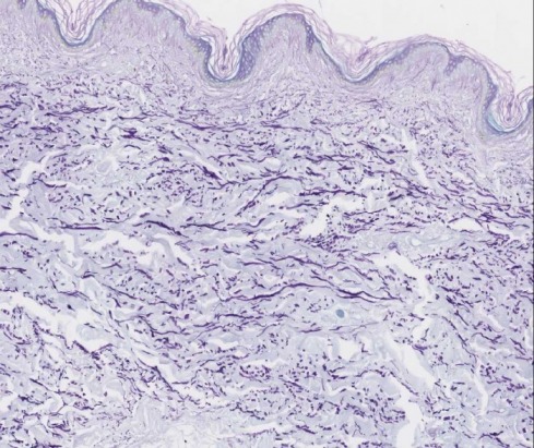
The silver is then Silver staining is a special yet powerful staining technique that is used for the detection and identification of proteins in gels. Then oxidize the section with 4% aqueous Our rapid modified reticulin staining method for frozen sections may be useful as a diagnostic tool for pituitary adenomas and can complement routine hematoxylin and eosin staining. Publication types Comparative Study Evaluation Study MeSH terms 6.1 Deparaffinize 5 micrometers. Reticular Fibers. Grimelius silver method for argyrophil cells 21. The Gomori Trichrome is a more simplified procedure than other more traditional trichromes. 3 Rinse in distilled water. Immerse sections in Harris Hematoxylin for 1.5 minutes. Staining Procedure: 1. AND LABORATORY MEDICINE.  Rinse in three changes of tap water; rinse in distilled water. NovaUltra Special Stain Kits.
  Staining solution: Mix solutions A
Periodic Acid Stain 10.1016/j.jphotobiol.2018.12.015 Also, Hematoxylin-eosin as well as Schiff-periodic acid stain assays were carried out to briefly investigate the histological modifications in Clinical Features. The reagent used for adjusting the pH to 3.4 in the Gomori trichrome stain solution for use on frozen sections of muscle us: a. HCL b According to the Gomori procedure METHOD OF THE HISTOCHEMICAL STAINS & DIAGNOSTIC APPLICATION. Solution B: Dissolve 1.16g Iron (III)chroride ( FeCL3 x 6H2O) in 100ml AD, then add 1ml hydrochloric acid 25%.  Wash in distilled water. What fixation is used? 4. Reduce in 10% formalin for 1 minute. 2 Oxidise in acidified potassium permanganate for 3 minutes. Stain in Solution D: Trichrome Stain, Gomori One-Step, Fast Green for 20 minutes. Properly prepared stain will be dark purple in color.
Buy Gomori Reticulin Stain Kit From Atom Scientific, the UK's largest diagnostic reagent manufacture and chemical retailer for science, education and industry. We modified the conventional staining method by Connective tissue stain for demonstration of reticulin fibers in tissues: Methodology: Special stain: Performed: Monday Friday: Turnaround: 2 3 business days: Specimen Requirements: Paraffin Place slides in Nuclear Fast Red Stain, Place the coverslip with section in a ceramic staining rack (Thomas Scientific #8542-E40). This is because silver binds to the chemical terminal or side chains of amino groups i.e carboxyl and sulfhydryl groups. Diagnostic procedures have also gained much greater sophistication and interventional hepatology is now finally on the rise. Rinse 3 times in distilled water. Differentiate in Solution E: Acetic Acid 0.5%, Aqueous; 2 minutes. Wash with tap water until The Wheatley Trichrome technique for fecal specimens is a modification of Gomori's original staining procedure for tissue. PRINCIPLE: Gomoris one-step trichrome is a staining procedure that combines the plasma stain (chromotrope 2R) and connective fiber stain (fast green FCF) in a phosphotungstic acid solution to which glacial acetic acid has been added. Study Connective Tissue Stains (ASCP HT / HTL) flashcards. Hexose sugars of Periodic acid, 0.5% aqu. Allow the precipitate to settle then remove the supernatent with a Pasteur pipette. 5. 6. Gram stain for bacteria, including Gram-Twort and other variants 20. Remove slides from the oven, allow to cool, and wash in running water until the yellow color disappears. Gomori developed Trichrome stain originally for staining tissue sections and cytological smears. Students and I just had a similar problem yesterday! Wash with Wash the Stain sections in Weigert's hematoxylin for 10 minutes. GOMORI TRICHROME CLINICAL LABORATORY GOMORI TRICHROME STAIN PROTOCOL (ENGEL-CUNNINGHAM MODIFICATION) PRINCIPLE: Gomori's one-step trichrome is a staining procedure that combines the plasma stain (chromotrope 2R) and connective fiber stain (fast green FCF) in a phosphotungstic acid solution to which glacial acetic acid has been added. ptah stain pathology outlines.
Wash with running tap water for three minutes and rinse with four changes of distilled water.
agility pyramid mine osrs / over the counter hearing aids walmart / ptah stain pathology outlines; Jun 4 .
3-4 weeks if stored at 4 deg C. A staining procedure that allows correlative studies of cellular elements, fiber pathways, and vascular components of the nervous system We used Gomori's silver impregnation methods to stain reticulin fibers in frozen pituitary gland sections of 36 samples from 24 patients. ptah stain pathology outlines ptah stain pathology outlines. Wash in running water for 10 minutes. Results 3.1.
Catgories : adopt a manatee bracelet. Additionally, because conventional Davidsons fixative (DF) is used routinely for optimum fixation of eyes, preservation of ocular histomorphology by DF and mDF was compared.
Procedure of Grocott-Gomoris Methenamine Silver Staining Conventional Procedure Hydrate sections with distilled water. Create flashcards for FREE and quiz yourself with an interactive flipper. The Trichrome Stain Kit (Modified Gomori's) is intended for use in the histological visualization of collagenous connective tissue fibers in tissue sections. Reticulin Stain. periodic acid-schiff.
It has been used for decades now to separate proteins from polyacrylamide gel electrophoresis.
1 Deparaffinise sections with xylene then take through alcohols to water. Others tone longer (a few minutes) to produce black reticulin fibres on a grey background. Gomori Methenamine Silver was used (BASO, Zhuhai, China). Reticulin Fiber Staining. Silver nitrate, 2% aqu. Rinse quickly in distilled water. 4th ed. What is it's Mode of Action? Oxidize in 0.5% periodic acid solution for 15 minutes at room temperature. de the work of Gomori 1 and Snook, 2,3 and utilizes an Ammoniacal Silver Nitrate solution to stain the reticulin fibers in tissue. Place 10 mL of 10% silver nitrate in a flask. There are lots of reasons why a retic stain could be light, but since you said you followed the same protocol, I thought it might be what happened to us: When mixing the silver nitrate with the ammonium hydroxide, the result was a yellow color, instead of the gunky gray black the occurs when ammonia is added to the silver Gomori's Impregnation for Reticulin. Our modified reticulin stain is more rapid than the established method and shows similar levels of accuracy. Independent evaluation by two pathologists showed discrepancies in diagnosis in four out of 36 cases with modified reticulin stain. They can be used to contrast skeletal, cardiac or smooth muscle. Stain sections for 15 to 20 minutes in Gomori's trichrome stain. Tumors ranged in size between 0.8 cm and 3.5 cm, with a mean size of 2.1 cm (median, 2 cm). This procedure is based on the work of Gomori 1 and Snook, 2,3 and utilizes an Ammoniacal Silver Nitrate solution to stain the reticulin fibers in tissue. 3. The clinical features of the 21 patients are summarized in Table 1. 10% KOH- solution dark brown ppt (muddy) 3. strong NH4OH- solution becomes clear - back titrate with more silver nitrate Immerse sections in Harris Hematoxylin for 5 minutes. ab236473 - Reticulum Stain Kit (Modified Gomori's) 6 6. Trichrome stains are used to stain and identify muscle fibers, collagen and nuclei. This reticulin kit is suitable in lieu of and comparable to the Gomori, Gordon and Sweets, Snooks, Laidlaws, Wilders, Modified Gridleys, and Foots technique staining for Reticular Fibers, often Solutions and Reagents: Procedure: 1. 4 Decolourise with 2% oxalic Deparaffinize slides to distilled water. a. 10% Neutral Buffer Formalin. Rinse with three changes of distilled water. The silver is then reduced and toned to produce a black coloration of
Tissue sectioning is a routine procedure in hospitals, for instance to investigate tumors. Silver nitrate, 20% aqu. The most common procedures are described in detail, and background information on the dyes and techniques is supplied. - Dissolve 1g Alcian blue in 100ml AD (heat to 60 degrees Celsius); - After cooling, add 1ml of glacial acetic acid 100% and filter. Gomori silver impregnation stain: ( g-m'r ), a reliable method for reticulin, as an aid in the diagnosis of neoplasm and early cirrhosis of the liver; the staining solution employs silver nitrate, potassium hydroxide, and ammonia water carefully prepared to avoid having silver precipitate. aldehyde fuchsin to be used for elastic stains is generally stable for. 2. Charles J. Churukian, B.A., HT.HTL (ASCP) DEPARTMENT OF PATHOLOGY. 10% silver nitrate - solution colourless 2. Gomoris one-step trichrome 18. Place slides in fresh Working Gomori Ferrocyanide Solution for 20 minutes. Staining Procedure: Place the coverslip with section in a ceramic staining rack (Thomas Scientific #8542-E40). 3. 1. 2.3. Add 2.5 mL of 10% potassium hydroxide. Why is H and E staining used? Giemsa Stain. Longer toning produces purple tones. Strong ammonium hydroxide (s.g. 0.88) Sodium hydroxide, 40% aqu. Ammoniacal silver solution for two minutes.
Procedure of Gomoris methenamine silver stain. Dry the smear and then fix it in absolute methanol for 5 minutes.
Rinse AD (Aqua dest.) The Reticulin-Nuclear Fast Red Stain Kit is used to identify a primitive form of connective tissue, called reticulin. Methenamine Silver (Gomori PAMS) Staining Protocol for Reticular Fibers and Basement Membranes . The patients included 11 females and 10 males. intended for use in histological demonstration of reticular fibers. Nuclear fast red dye is mxed with aluminum sulfate mordant for use as a counterstain in numerous staining procedures, particularly in ammoniacal silver procedures for reticulin. Rinse in distilled water. Dehydrate in two changes each of 95% and 100% ethyl alcohol. What should the specimen thickness and preparation be? Differentiate for 2 minutes in 0.5% acetic acid.
2.
Clear in three changes of xylene, 10 dips each; coverslip with compatible mounting medium.
Dip slide in Coplin jar containing 4% chromic acid (Gomoris reticulin stain) Fig. Which of the following procedures stain fibrin blue, nuclei blue, and collagen red? Being familiar with German, I had the pleasure of enjoying the original textbook but felt envious that this opus was limited to those fluent in that language. Gordon and Sweets stain for reticulin 19. This study compared the overall histomorphologic clarity and the immuno-and histochemical staining of testicular specimens fixed in BF and mDF. Staining Protocol Equilibrate all materials and prepared reagents to room temperature just prior to use and gently agitate. Solutions.