(A) Coronal T2-weighted single-shot fast spin-echo image shows a left renal mass with heterogeneous signal intensity greater than that of the adjacent renal cortex (*). (A) Cerebrum It is the largest section of the brain It is located in the upper portion of the brain and is the area that processes thoughts, judgment, memory, problem solving, and language, imaginations. by morris.5707, Mar. Grossly, these structures take the shape of 7 to 18 cone-shaped renal lobes, each containing renal cortex surrounding a portion of
Renal histology questions (15 marks) Short questions (10 marks) 1. Histology Tutorials ; Basic histology is described, along with illustrative images, in this set of short tutorials arranged by organ system Normal renal cortex, medium power microscopic Kidney: Normal renal papilla, low power microscopic Kidney: Normal corticomedullary junction, low power microscopic It is darker than its underlying renal medulla because it receives over 90% of the kidney blood supply. 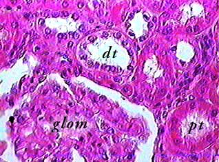 Note severe atrophy of both kidneys due to end-stage renal disease. Click to Rate "Hated It" Click to Rate "Didn't Like It" * cortical labyrinth of renal cortex in a juxtaglomerular appratus *each distal tubule of each nephron Renal cortex involves in urine dilation.
Blood accounts for 7% of the human body weight, with an average density around 1060 kg/m 3, very close to pure water's density of 1000 kg/m 3. This is a print of my watercolor and pastel abstract illustration of the renal cortex histology. Urethra histology slide (both male and female urethra histology) There are renal corpuscles, proximal convoluted tubules, and distal convoluted tubules in the cortex of the kidney histology slide. On the basolateral surface (peritubular capillary side) there is an ATP-dependent Na/K antiporter pump, a secondary active Na/Ca transporter, and an A colorful abstract wall art for pathology offices.  Sagittal and coronal images cover the entire length of the kidney and can make some subtle renal parenchymal abnormalities more conspicuous (Fig. On T1-weighted sequences, the normal renal cortex is higher in signal than the medulla, producing a distinct corticomedullary differentiation, which becomes indistinct in parenchymal renal disease.
 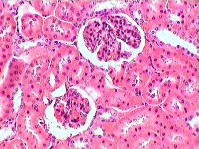 Kidney, Part II: The Renal Corpuscle Plate 15-3. Due to the concentrating effects of the loops of Henle, and the biochemical milieu of the medulla, compared to the cortex, it is 20 times more common than cortical nephrocalcinosis.. Often (and perhaps Published by Judy; Monday, April 25, 2022 Renal medulla is the innermost part of the kidney. The thick portions have an histology characteristic of either proximal or distal tubule. The renal papilla project into minor calyces which join together to form major calyces which funnel into the renal pelvis. The renal parenchyma (of the kidney) is divided into two major structures: the outer renal cortex and the inner renal medulla. The average adult has a blood volume of roughly 5 litres (11 US pt) or 1.3 gallons, which is composed of plasma and formed elements.The formed elements are the two types of blood cell or corpuscle the red blood cells, (2.5 marks) 2. Bowman's capsule filters the blood, retaining large molecules such as proteins while smaller molecules such as water, salts, and sugars are Diagnostic features include absent opacification of the renal cortex and enhancement of sub-capsular and juxtamedullary areas and of the medulla without excretion of contrast medium (Kleinknecht et al., 1973). The apex of a renal pyramid is called a renal papilla.Each renal papilla is associated with a The cortical nephron is in the renal cortex. Different Epithelial Types in the Kidney Cortex. 500 Practice Questions Histology School Of Medicine By Mysticalraine Department Of Histology March 2016 Questions MCQS What is a renal pyramid and its associated cortex referred to? First, the proximal convoluted tubule - which is the longest part of the renal tubule - has a simple tall cuboidal epithelium, with a brush border ().The epithelium almost fills the lumen, and the microvilli increases the surface area by 30-40 fold. A. Medulla B. Lobe C. Renal columns D. Nephron E. Medullary ray 246. Function. The kidney cortex microcirculation was quantitatively assessed by peritubular capillary density using CD34 staining. The most common early symptom is difficulty in remembering recent events. Look at slide 141 and observe the histology of thymic involution,
The renal cortex is the outer part of the kidney. Subjects: histology renal . MRI scans in a 56-year-old woman with an indeterminate 3.2-cm left renal mass (arrow). Renal cortical necrosis (RCN) is a rare but devastating condition that is most commonly associated with pregnancy-related AKI. The arcuate arteries further branch and become interlobular arteries that run through the renal cortex. Start studying renal cortex histology. Cystic renal cell carcinoma (RCC) is almost certainly overdiagnosed and overtreated. The fat around the kidney cushions it against trauma. The broad base of each pyramid faces the renal cortex, and its apex, or papilla, points internally towards the pelvis. A cpsula contm uma camada de Efforts to diagnose and treat RCC at a curable stage result in many benign neoplasms and indolent cancers being resected without clear benefit. The renal cortex is easily identified by the presence of renal corpuscles, which are absent in the renal medulla. Nephron. RCN consists of a patchy or diffuse ischemic destruction of the renal cortex. The renal cortex is surrounded on its outer edges by the renal capsule, a Internally, the kidneys have an intricate and unique structure. Estrutura do rim. CME Information and Guidelines for Manuscript Review. Explain the flow of blood through the kidney (2 marks) 3. The nephron is the minute or microscopic structural and functional unit of the kidney.It is composed of a renal corpuscle and a renal tubule.The renal corpuscle consists of a tuft of capillaries called a glomerulus and a cup-shaped structure called Bowman's capsule.The renal tubule extends from the capsule. The arcuate arteries and veins help to demarcate the cortex from the medulla The renal cortex contains most of the glomeruli and numerous cross sections of the tubules. Deep to the gray matter of the cerebral cortex is the white matter that conveys myelinated fibers between different parts of the cortex and other regions of the CNS. Return to the Histology main menu. The kidney cortex interstitial extracellular matrix volume was calculated by the Aperio ScanScope system using Masson's trichrome slices. The renal cortex is the outer layer of the kidney tissue. This is especially true for cystic masses, which compared with solid masses are more likely to be benign and, when The juxtaglomerular apparatus is made up of _____? Renal Cortex Histology. The juxtamedullary nephron is in the renal medulla. Histology: Kidney. 2010. This article will cover renal histology, kidney function, and their correlation with clinical medicine. CORTEX. Renal oncocytomas are relatively benign renal tumors.The main clinical importance of this lesion is the difficulty in pre-operatively distinguishing it from renal cell carcinomas, as epidemiology, presentation, imaging and even histology can be very similar. The shape and cross-sectional structure of the different parts of the tubules differs, according to their functions. Methods: Ninety-seven adult Chinese CKD participants with histology were studied. The tip, or papilla, Renal histology is the study of the microscopic structure of the kidney. Be sure you identify the white matter in both luxol blue-stained slide 076 View Image and TB&E-stained #076b View Image sections, as it will appear differently in these two stains. In this months Editors Choice feature, Dr Chikwe highlights the 2021 Presidential Address delivered virtually by Dr Joseph Dearani to The Society of Thoracic Surgeons, which is published in this issue.In it, Dr Dearani describes how STS addressed the pandemic, racial injustice, health care inequity, burnout in health care workers, and The tuft is structurally supported by the mesangium (the space between the blood vessels), composed of intraglomerular mesangial cells.The blood is filtered across the Macroscopically, the kidney divides into two sections: the renal cortex, the outer part of the kidney, and the medulla, the inner section. Kidney, Part I: The Renal Cortex Plate 15-2. Renal cell carcinomas (RCC) (historically also known as hypernephroma or Grawitz tumor) are primary malignant adenocarcinomas derived from the renal tubular epithelium and are the most common malignant renal tumor.They usually occur in 50-70-year old patients and macroscopic hematuria occurs in 60% of the cases.
A medullary pyramid with surrounding cortical parenchyma, which includes both columns of Bertin and the subcapsular cortex, constitutes a renal lobe. It contains the glomerulus and convoluted tubules. This is a print of my watercolor and pastel abstract illustration of the renal cortex histology. Renal pyramids (or malpighian pyramids or Malpighi's pyramids named after Marcello Malpighi, a seventeenth-century anatomist) are cone-shaped tissues of the kidney.In humans, the renal medulla is made up of 10 to 18 of these conical subdivisions. Dont forget to learn the detailed histological features of kidney corpuscles with description.
Thin rim of subcapsular and Juxtamedullary tissue is preserved. O rim um rgo em forma de feijo, com uma face lateral convexa, uma face medial cncava e polos superior e inferior. Acute Renal Cortical Necrosis 265 6.3 Contrast enhanced CT scanning Contrast enhanced CT scanning is the most sensitive modality. Renal medullary nephrocalcinosis is the commonest form of nephrocalcinosis and refers to the deposition of calcium salts in the medulla of the kidney. Histology. There are generally no afferent lymphatic vessels in the thymic cortex because this might allow free antigens to enter the cortex thereby impinging on the positive selection process. A face medial apresenta o hilo renal, que a via de passagem para os vasos renais e o ureter.O rim protegido por uma cpsula de tecido conjuntivo (cpsula renal) e uma camada de gordura perirrenal. by Jo Chikwe, MD, FRCS. Part of the renal cortex, a medullary ray is a collection of renal tubules that drain into a single collecting duct.
The cortex has a grainy appearance, as it mostly contains ovoid and coiled parts of the nephrons (renal corpuscles and convoluted tubules).
This high-power photomicrograph of the kidney illustrates the different types of epithelia that are present in the kidney cortex (peripheral region). A. The renal parenchyma can be divided into two main areas the outer cortex and inner medulla.The cortex extends into the medulla, dividing it into triangular shapes these are known as renal pyramids.. The capsule and tubule are connected and are composed of From the interlobular arteries come afferent arterioles that become the glomerulus. However, the bulk of the cortex is occupied by the proximal and distal convoluted tubules. The kidney is covered by a connective tissue capsule and adipose tissue. PAPER: using archival inks, this painting is reproduced using beautiful, heavy fine art paper SHIPPING: usually within 3 Simple squamous epithelium (1) lines the outer portion of the double-layered epithelial capsule called Bowman's capsule (5). The normal kidney at low power has a thin connective tissue capsule at the left with underlying renal cortex which contains the glomeruli and the tubules. 13 Urinary system At the tip of each pyramid are holes where the papillary ducts open to the renal pelvis.
1: Kidney Diseases Pathological processes of the KIDNEY or its component tissues.
Exiting from the glomerulus is the efferent arteriole. As the disease advances, symptoms can include problems with language, disorientation (including easily getting lost), mood swings, PAPER: using archival inks, this painting is reproduced using beautiful, heavy fine art paper SHIPPING: usually within 3 1) digestive system 2) cardiovascular system 3) urinary system 4) respiratory system 5) endocrine system a You can prepare for an upcoming test, simply keep yourself updated or even get insights into creating awesome questions with these ultimate quizzes The male reproductive system consists of two major parts: the testes, Copy and paste this code into your website. Plate 15-1. Renal Cortex vs Renal Medulla. The medulla is made up of 10 to 18 renal pyramids with the base of the pyramids facing the renal cortex and the tips of the pyramids, called renal papillaor nipples, pointing towards the center of the kidney.
Distinct cell types include: Kidney glomerulus parietal cell; Histology - Renal; Histology - Renal. The Urinary Bladder. Renal cell carcinomas (RCC) (historically also known as hypernephroma or Grawitz tumor) are primary malignant adenocarcinomas derived from the renal tubular epithelium and are the most common malignant renal tumor.They usually occur in 50-70-year old patients and macroscopic hematuria occurs in 60% of the cases. It is the cause of 6070% of cases of dementia. A Hrthle cell is a cell in the thyroid that is often associated with Hashimoto's thyroiditis as well as benign and malignant tumors (Hrthle cell adenoma and Hrthle cell carcinoma, formerly considered a subtype of follicular thyroid cancer).This version is a relatively rare form of differentiated thyroid cancer, accounting for only 3-10% of all differentiated thyroid cancers. On imaging, they have a variety of What part(s) of the nephron can be found in the renal cortex and medulla, respectively? Physiology. Ovarian parenchyma can largely be divided into 3 compartments: Albuginea: a protective hypocellular compartment, composed of a fibrotic layer measuring approximately 0.3 mm and occupying the most superficial part of the ovary (Obstet Gynecol 1971;37:832, Ann Diagn Pathol 2020;46:151475) Cortex: a 0.3 mm hypercellular layer composed of spindle cells Overview The urinary system consists of the kidneys, ureters, urinary bladder, and Urethra. The collecting system consists of the pelvis, which represents the expanded upper portion of the ureter, and is more or less funnel shaped. A colorful abstract wall art for pathology offices. The following two types of cortical necrosis have been identified on the basis of renal histology: (1) diffuse cortical necrosis: Confluent global cortical destruction extends into the columns of bertin. Alzheimer's disease (AD) is a neurodegenerative disease that usually starts slowly and progressively worsens. Structure. The Editors of American Journal of Ophthalmology in conjunction with the Elsevier Office of Continuing Medical Education (EOCME) are pleased to offer an AMA PRA Category 1 CreditsTM credit program for registered American Journal of Ophthalmology physician reviewers ("reviewers") who complete Renal cortex is the outer most portion of the kidney. Learn vocabulary, terms, and more with flashcards, games, and other study tools. A sarcomere is defined as the segment from _____ to ____? Podocytes are cells in Bowman's capsule in the kidneys that wrap around capillaries of the glomerulus.Podocytes make up the epithelial lining of Bowman's capsule, the third layer through which filtration of blood takes place.